cNeuro® cMRI
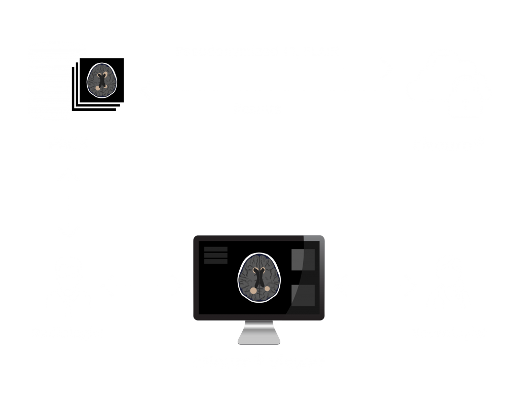
cMRI provides fully automated brain MRI quantification for increased throughput, greater objectivity, and high-quality reporting for improved patient care.
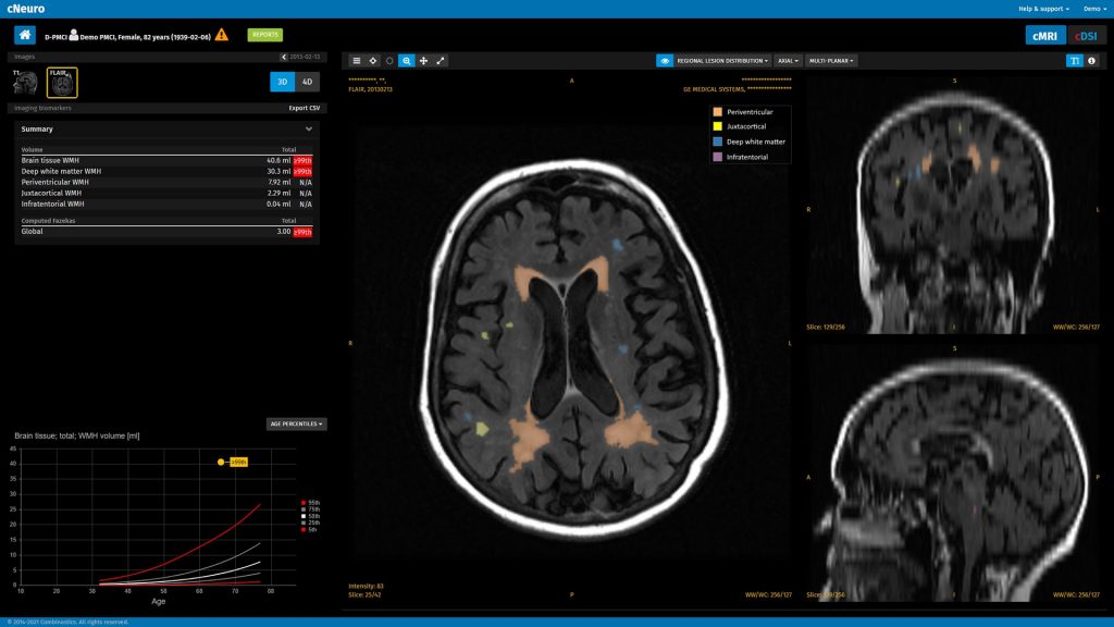
cNeuro® cMRI, our cloud-based AI software, integrates with PACS, and results can be viewed in PACS or using the cNeuro browser-based viewer.
Radiologists benefit from increased accuracy and sensitivity, less time spent per read, and help managing complex patients in collaboration with referring clinicians.
Benefit from objective data
Objectively quantify disease-specific abnormalities and patterns and reduce the variability in radiological interpretation.
Fully automated, reliable, reproducible analysis of brain T1 and FLAIR MRI:
- Volumes of 133 cortical and subcortical regions (T1)
- White-matter lesions
- Additional disease specific biomarkers
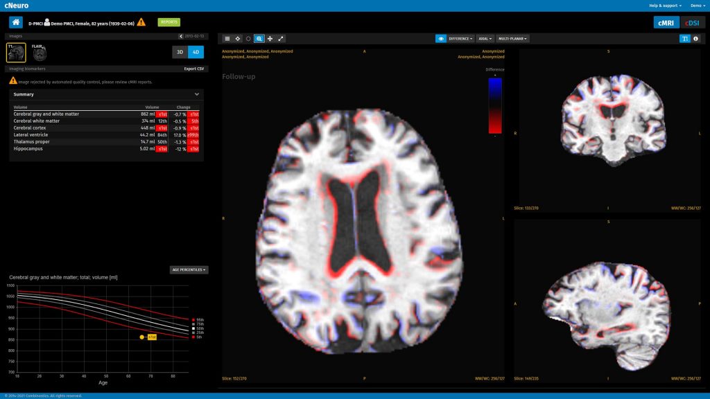
- Quantification of longitudinal changes in brain structure and lesions
Statistical comparison of results to normative reference data adjusted for age, sex, and intracranial volume with results presented in tabular form or as age-corrected and gender-corrected plots
Interactive image review, with the ability to compare multiple timepoints and toggle overlays on/off for easy assessment of segmentation results
Watch a demo of cMRI
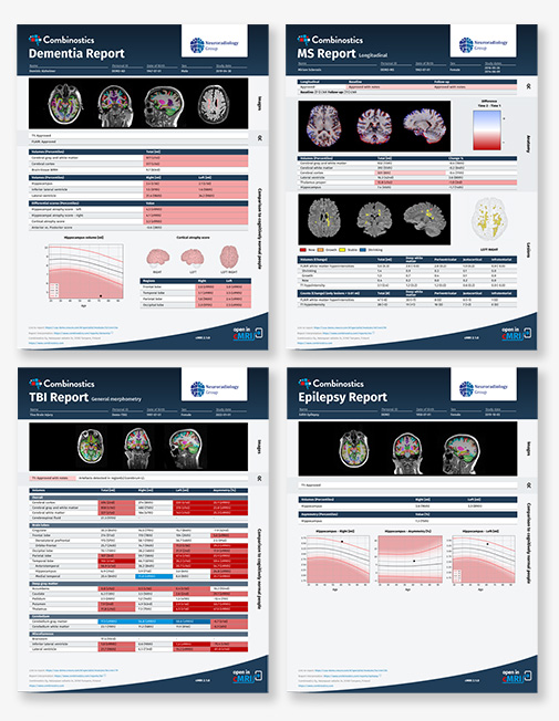
Improve collaboration and referrals with clinically focused reports
Improve collaboration, provide greater value, reduce image-related queries, and drive more referrals through streamlined, high-quality, consistent communication with referring clinicians.
Our new clinically focused cMRI reports for dementia, multiple sclerosis (MS), traumatic brain injury (TBI), and epilepsy facilitate patient management as well as communication between radiologists, neurologists, patients, and their caregivers.
Regulatory Compliance
CE marked, FDA 510(k) cleared, and ANVISA (Brazil)
Security
- All data transfer uses SSL encryption
- Stored data are anonymized and encrypted
- More information is available in a separate security statement.
Indications for Use
cNeuro cMRI is intended for automatic labeling, quantification and visualization of segmentable brain structures from a set of MR images. The software is intended to automate the current manual process of identifying, labeling, and quantifying the segmentable brain structures identified on MR images. The intended user profile covers medical professionals who work with medical imaging. The intended operational environment is an office-like environment with a computer.
The cMRI compute volumes of brain structures were validated for accuracy and reproducibility with
1350
participants.


